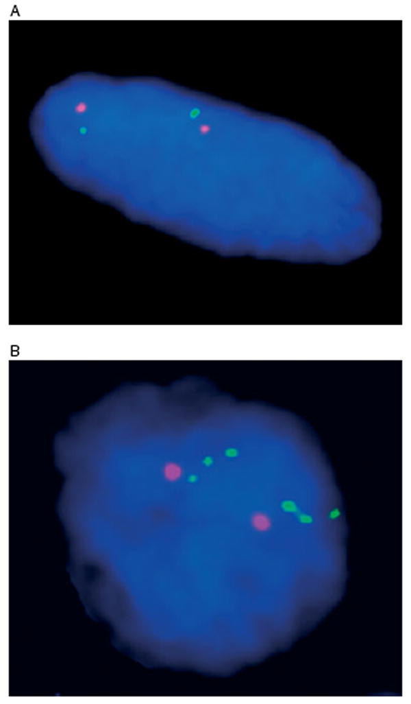
Figure 3.

(A) FISH analysis of abdominal tumour 1 (spindle cell morphology) from patient 8, with disomic (FISH-negative) pattern. The chromosome 4 centromere probe is shown in orange and the KIT probe in green. (B) FISH analysis of stomach wall (2) tumour (epithelioid morphology) from patient 10, with KIT amplification. Chromosome 4 centromere probe is shown in orange and the KIT probe in green
