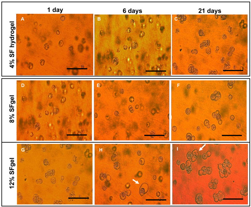
Figure 6.
Microscopic imaging of hMSCs encapsulated and cultured in silk fibroin hydrogels at 4, 8, and 12% (w/v). Microscopic images were taken at day 1 (A, D, G for 4, 8, and 12%, respectively), 6 (B, E, H for 4, 8, and 12%, respectively), and 14 (C, F, I for 4, 8, and 12%, respectively). For the 4% gel, hMSCs retained round shape morphology and were nonaggregated in the gel. For the 8 and 12% gel, cells largely deformed and aggregated by day 21, especially for the cells in the 12% gel, as indicated by the arrows in H and I. Bar = 100 μm in all images.

