Figure 7.
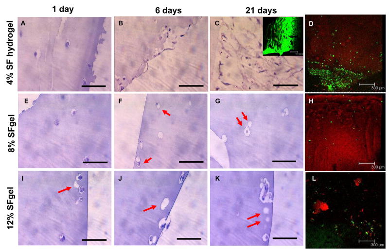
Histological and cell live-dead analysis of hMSCs encapsulated and cultured in silk fibroin hydrogels at 4, 8, and 12% (w/v). The gel samples were subjected to histological evaluation (H&E) at day 1 (A, E, I for 4, 8, and 12%, respectively), 6 (B, F, J for 4, 8, and 12%, respectively), and 21 (C, G, K for 4, 8, and 12%, respectively), and cell live and dead assay at day 21 (inset of C and D for 4%, H and L for 8 and 12%, respectively). For the 4% gel, hMSCs either retained a round shape in the gel or changed to spindle-like shape and proliferated near the gel surface (A–C), and most cells were alive near the gel surface (inset of C) and in the gel (D), as determined by the dominant green fluorescence. For the 8 and 12% gels, hMSCs largely changed morphology and many of them died, aggregated and/or dissolved, as seen by empty cavities in histological images (arrows in F, G, I, J, K) and few green fluorescent spots in the live-dead assay (H, L). Bar = 100 μm in A–C, E–G, I – K; 101.2 μm in the inset of C; 300 μm in D, H, L.

