
Fig. 10.
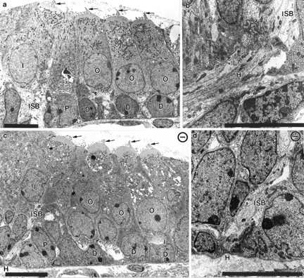
Transmission electron microscopic images of the organ of Corti in newborn control and mutant animals. Electron micrographs show the organ of Corti with the three rows of outer hair cells (O), Deiter’s cells (D), one row of inner hair cells (I), and pilar cells (P) in control (a) and NT-3 mutant (c) animals. Other than the plane of the section, which is slightly oblique in c, there are no differences between the control (a) and the NT-3 mutant littermate (c). Note the presence of apical specializations in all hair cells (arrows in a, c). The habenula perforata (H) has no fibers passing through at the basal turn of NT-3 mutant mice (c, d) but has numerous fibers joining the inner spiral bundle (ISB) underneath the inner hair cell in control animals (a, b). Scale bar, 10 μm in each panel.