
Fig. 4.
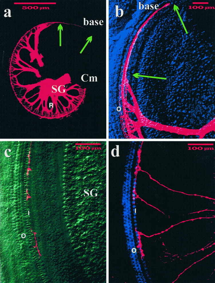
Distribution of sensory afferent fibers in the basal turn of P0 NT-3 mutant homozygotes. All afferents (a, b) and subsets of afferents (c, d) were filled with DiI from the brainstem. Note a that the basal turn of the cochlea (base) lacks spiral ganglion cells (SG) and radial fibers (R), both of which are present in the middle turn (Cm). Nevertheless, fibers extend along inner hair cells toward the base (arrows). As illustrated b, few fibers extend to outer hair cells (O) near the middle turn. Selective labeling reveals the distribution of individual afferents in the middle basal turn (c, d). Note that the terminal arbor of each afferent fiber is restricted to several inner hair cells (I) with no processes crossing to the three rows of outer hair cells (O), which appear normally developed in this differential interference contrast (c) or fluorescence image (d). Scale bar, 500 μm for all images.