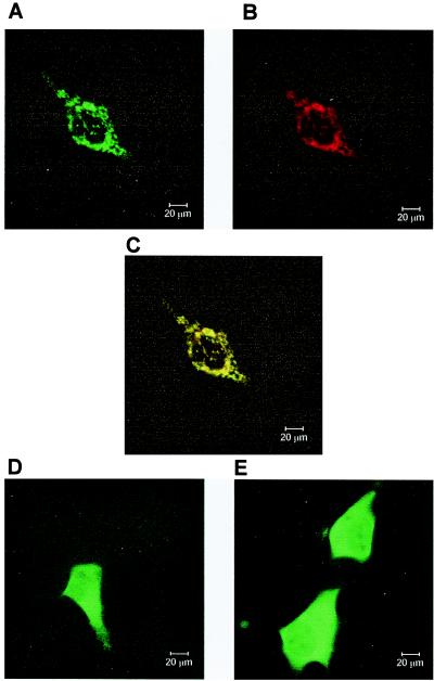
Figure 5.
Mitochondrial localization of dNT-2 shown by laser confocal microscopy. 293-2-100 cells were transfected with ecdyson-inducible vectors expressing the deoxyribonucleotidases as GFP-fusion proteins. (A–C) Fluorescence of the identical cell overproducing full-length dNT-2. (A) GFP fluorescence; (B) fluorescence of mitochondria stained with tetramethylrhodamine methyl ester; (C) overlay of A and B showing colocalization of dNT-2 and mitochondria. (D) GFP fluorescence of cells overexpressing dNT-2 without mitochondrial leader. (E) GFP fluorescence of cells overexpressing dNT-1.

