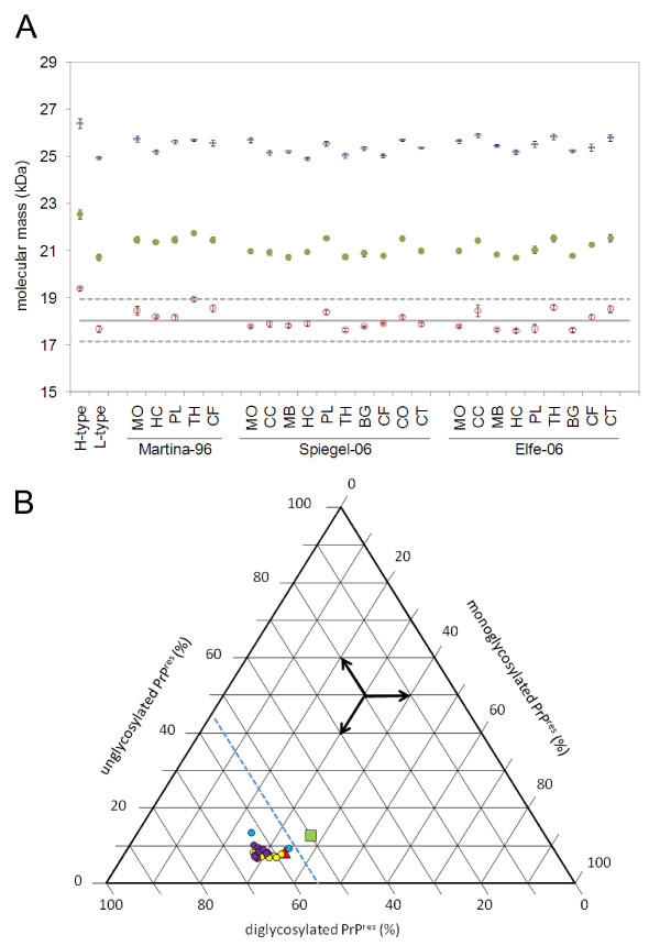
Figure 3.
Biochemical PrPres typing in different brain regions of selected aged cattle with C-type BSE. All analyses were conducted as described for figure 2. a) Molecular masses of unglycosylated (red open squares), monoglycosylated (green filled circles) and diglycosylated (blue rectangles) PrPres and b) relative intensities of the un-, mono- and diglycosylated PrPres moieties of the C-type BSE cases Spiegel-06 (yellow circles), Martina-96 (blue circles) and Elfe-06 (violet circles) as compared to L-type BSE (green square) and H-type BSE (red triangle). Whenever available, the following brain regions were analyzed: MO, medulla oblongata; CC, cerebellar cortex; MB, midbrain; HC, hippocampus; PL, piriform lobe; TH, thalamus; BG, basal ganglia; CF, frontal cortex; CO, occipital cortex; CT, temporal cortex.

