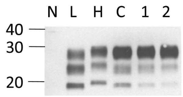
Figure 4.
Western immunoblot (protocol II) analysis of two ambiguous samples with mAb Sha31. Samples from cases with BSE Martina-96 (lane 1, thalamus) and Bunaug-02 (lane 2, medulla oblongata) compared to L-type BSE (L), H-type BSE (Charly-04; H) and C-type BSE (C). Note that the unglycosylated PrPres in both samples migrates in line with that in C-type BSE, and different from that in H- type BSE. On the left, a molecular mass marker is indicated in kDa.

