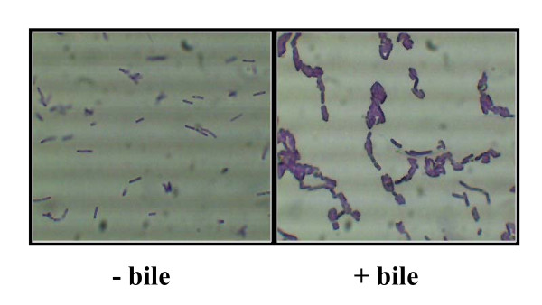
Figure 3.
Microscopy of cells cultured in bile. L. monocytogenes cells were prepared exactly as for biofilm assays (i.e. grown in BHI alone (- bile) and BHI supplemented with 0.3% oxgall (+ bile) and subsequently washed in 1/4 strength Ringers solution). 10 μl of crystal violet was added to 100 μl washed cells in a microcentrifuge tube and mixed. 10 μl was spotted onto slides and viewed with a Leica DMLS microscope containing a digital eyepiece. Three random fields were viewed and representative images were captured.

