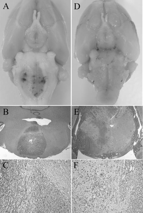
Figure 4. Representative photographs and microscopies of brainstem gliomas in adult (A, B, and C) and young (D, E, and F) rats.
A. Depicted is the relatively focal lesion with a displacement of long tract in an adult rat harboring brainstem tumor. B and C: Hematoxylin and eosin stain of brainstem cross section of an adult rat revealed highly cellular lesions invading the white matter with a relatively clear boundary. Abundant necrosis was consistently observed (B, magnification ×1; C, magnification ×10). D. Depicted is the enlargement of the entire brainstem without focal lesion in a young rat harboring brainstem tumor. E and F: Hematoxylin and eosin stain of brainstem cross section of a young rat revealed more infiltrative and diffuse lesion, with a high degree of brainstem parenchyma invasion. Endothelial proliferation was evident and angiogenesis were frequently observed (E, magnification ×1; F, magnification, ×10).

