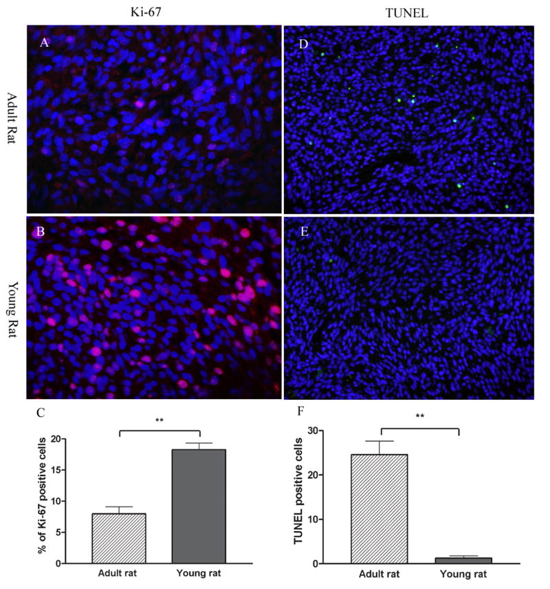
Figure 5. Tumor proliferation and apoptosis in adult (A, B and C) and young (D, E and F) brainstem gliomas.
Tumor cell proliferation was determined by immunohistochemistry of Ki-67 protein in red (A, B). Nuclei were counter stained by DAPI in blue. Ki-67 index of brainstem gliomas in young rats were significant higher than that of adult rats (C). Apoptosis was determined by TUNEL staining in green (D, E). Nuclei were counter stained by DAPI in blue. Few TUNEL positive cells were observed in the brainstem gliomas of young rats. While, a number of tumor cell underwent apoptosis in adult rats (F). Significant differences between the two groups were indicated by ** P<0.001.

