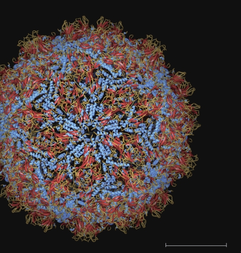
Figure 4.
View of the PBV particle down an I5 axis. The individual subunits are coloured according to secondary structure elements: α-helices blue, β-sheets red and random coil orange/light brown. Note the I5 loop with the conserved 157-NSG-159 segment at the centre, forming a diaphragm-like structure at the I5 axis. Bar: 100 Å

