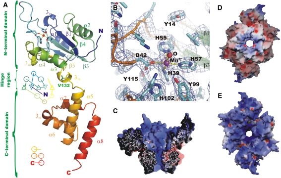
Figure 2.
Structure of the RepB monomer, active site and electrostatic surface representations. (A) A cartoon drawing of a single protomer of RepB, indicating the topology and secondary structure elements (Kabsch and Sander, 1983) of the OD and OBD domains. V132, located in the hinge region between the two domains, is shown in green, the active site is indicated using a ball-and-stick representation of the metal and its first coordination sphere. (B) A close-up of the active site, including the σA-weighted 2Fo−Fc electron density map contoured at 1.2σ (blue) and 4σ (purple). The metal-binding residues and the coordinated water/hydroxide ligand (‘O') are indicated. (C) The electrostatic potential on the solvent-accessible surface of a cross-section of the C3-structure and of the C2-hexamer, viewed (D) from the C-termini and (E) from the N-termini. Blue, red and white represent positively charged, negatively charged and neutral surface, respectively.

