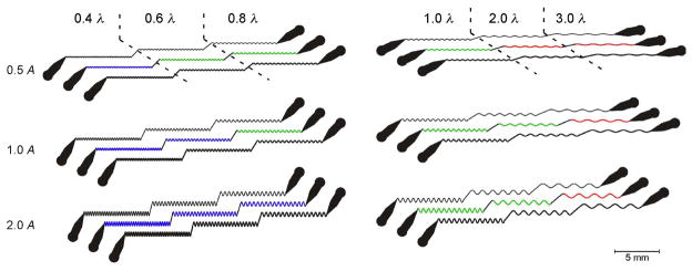
Figure 5.
Diagram of the waveform sampler device. The device consists of sinusoidal channels grouped into six triplets. Within a triplet, a single design is replicated with channels widths of 60, 80, and 100 μm (top to bottom). Each design comprises three sinusoidal domains connected by straight segments. Each domain has a unique combination of wavelength and amplitude (peak-to-peak), as indicated by the scale factors in the margins. The scale factors are nominally wild-type values of amplitude (A = 200 μm) and wavelength (λ = 500 μm). Domain color represents qualitatively distinct behaviors of worms in the channels: green, crawling with a well controlled waveform; blue, crawling with a partly controlled wave form; red, no crawling.

