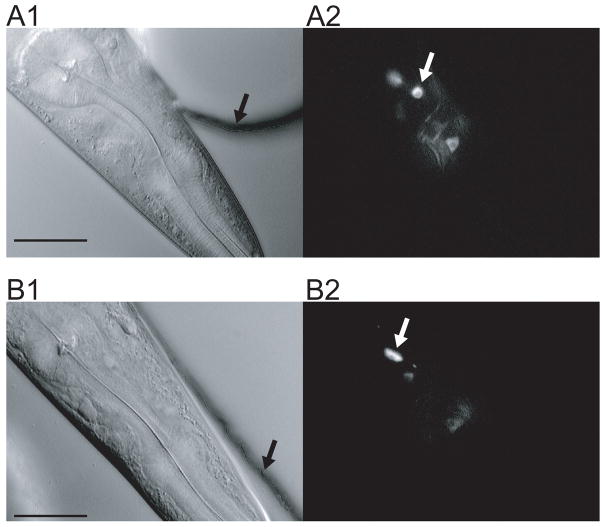
Figure 7.
Neuronal cell bodies imaged in the artificial soil and waveform sampler devices. A. Artificial soil. The worm was confined in the device shown in Fig. 3C. The black arrow indicates the wall of a post; the white arrow indicates a neuron cell body. B. Waveform sampler. The worm was confined in the channel shown in Fig. 6A. Arrows have the same meaning as in A. Left, differential interference contrast (DIC) image. Right, fluorescence image. Images were taken using Zeiss Axioskop FS equipped with a video camera (Dage MTI VE1000, Michigan City, IN). Fluorescence images are averages of five frames (29.98 frames/sec). Scale bar, 30 μm.

