
Figure 3. RanBP2 Binds to Topo IIα in Mitosis and Is Essential for Its Accumulation at Inner Centromeres.
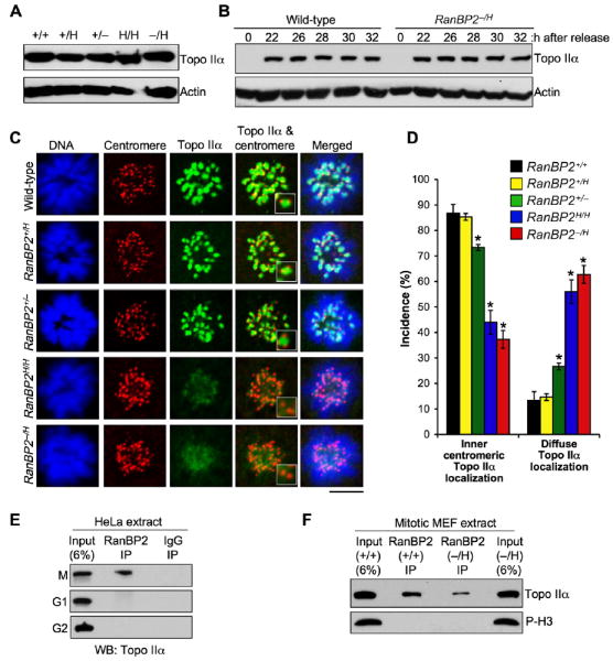
(A) Western blots of asynchronous MEF lysates probed with antibody to Topo IIα. Actin served as a loading control.
(B) Western blots of synchronized RanBP2+/+ and RanBP2−/H MEF lysates. MEFs were synchronized in G0 by serum starvation and then released for the indicated durations in serum-containing medium. Nocodazole was added 23 hr after cells were released. Blots were probed for Topo IIα. Actin was used as loading control.
(C) Immunolocalization of Topo IIα in MEFs with various levels of RanBP2 during prometaphase. Centromeres were visualized with ACA antibody. DNA was stained with Hoechst. Magnified images of the centromeric and inner centromeric regions are shown in the insets. Bar = 10 μm.
(D) Quantification of prometaphases with inner centromeric versus diffuse localization of Topo IIα. Seventy-five prometaphases were analyzed per genotype (three independent MEF lines were analyzed per genotype). Error bars indicate SEM, *p < 0.05 versus wild-type cells (Chi-square test).
(E) Immunoblots of mitotic (M), G1, or G2 HeLa extracts subjected to immunoprecipitation with RanBP2 antibody and analyzed for coprecipitation of Topo IIα.
(F) Immunoblots of mitotic extracts from RanBP2+/+ and RanBP2−/H MEFs subjected to immunoprecipitation with RanBP2 antibody and analyzed for coimmunoprecipitation of Topo IIα. Phosphohistone H3 (P-H3) signals indicate that similar amounts of mitotic cells were present in the lysates used for immunoprecipitation.