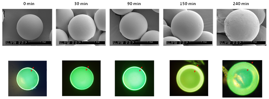
Figure 1.
Scanning electron microscopy (SEM) (upper panels) and fluorescent microscopy (lower panels) of sliced HTSC beads prepared at 80°C in MeOH (5mL) under various polymerization time. The red arrows marked the thickness of the hydrogel layer of the shell-core bead. The bead interior was capped with Boc. The outer layer was labeled with FITC.

