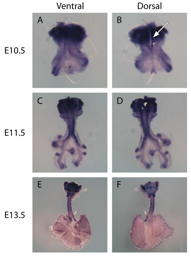
Figure 1.

Whole mount in situ hybridization of Larynx-trachea-lung explants from E10.5 (A & B), E11.5 (C & D), and E13.5 (E & F) using RNA probes to Col2A1. Both ventral (A, C, E) and dorsal (B, D, F) views are presented. Col2A1 is expressed specifically by chondrocytes, and therefore binding of the Col2A1 anti-sense RNA probe denoted by blue staining identifies chondrocytes in the developing upper respiratory tract. At E10.5, the upper respiratory tract begins as a simple out-pouching of the ventral foregut endoderm. At this stage of development there is a cleft along the posterior aspect of the trachea from the level of the larynx to the carina (see arrow, figure 1B). To form the mature configuration of the thyroid, arytenoid, cricoid, and tracheal cartilage, the chondrocytes present at E10.5 must expand directionally. Col2A1 expression is also present in the distal aspects of the lung where it is known to be expressed in the endodermal compartment. Sense probes used to verify non-specific binding, showed no stain (data not shown). (A & B were taken at 10X, C & D at 5X, and E & F at 2.5X)
