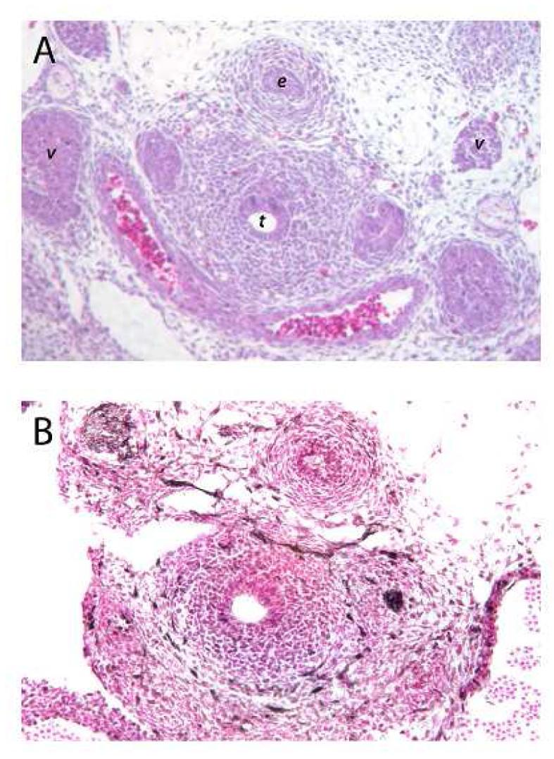
Figure 2.

(A) H&E stained axial section through the mid trachea of an E12.5 mouse embryo. The tracheal lumen (t) and surrounding mesenchymal cells are readily apparent. Posterior to the trachea is the esophageal lumen (e). Lateral to the trachea are the lumen of vascular structures (v). (B) Antibodies to ERK highlight (dark brown stain) cells at the periphery of the mesenchymal cells surrounding the trachea. ERK positive cells are also seen in the mesenchymal cells surrounding the esophagus, and adjacent vascular structures. (This section of tissue does have a tear artifact on the left side between the trachea and adjacent vascular structure. Controls with secondary antibody alone showed not staining (data not shown). (Figures are taken at 20X)
