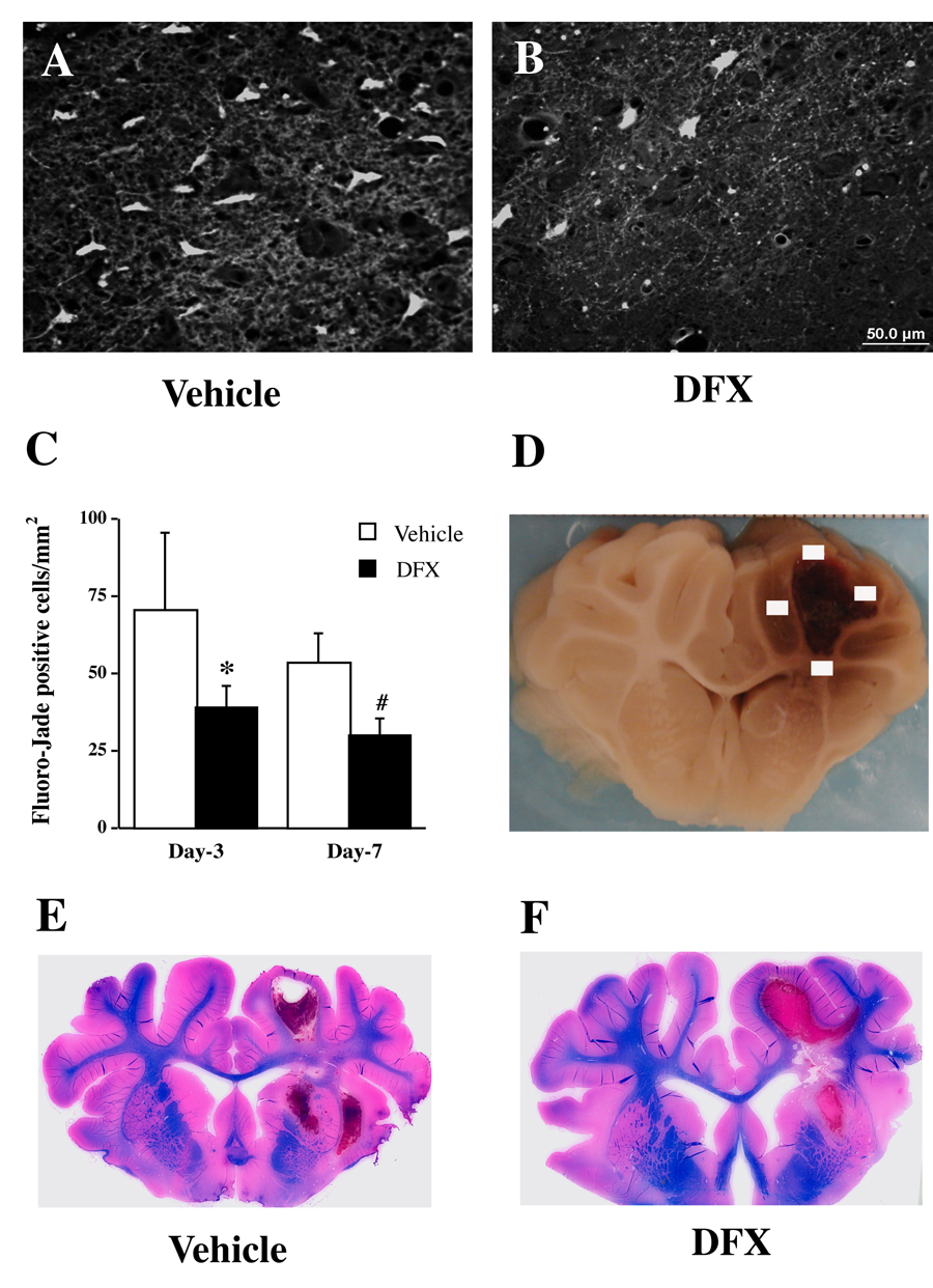
Figure 3.
Fluoro-Jade C positive cells in the perihematomal area (A–C) and Luxol fast blue staining (E & F) after ICH. Part D shows four sampled fields for Fluoro-Jade C cell counting. Pigs had ICH and were treated with either vehicle or deferoxamine. Values are means±SD, n=4, *p<0.05, #p<0.01 vs. vehicle. Scale bar (A & B)=50 µm.

