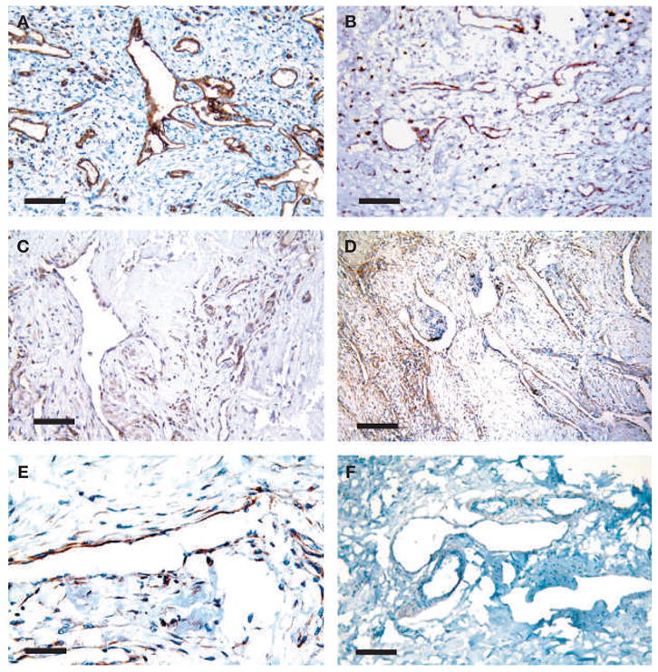
Figure 3.

Immunohistochemical analysis of the patient’s tumor tissue. (A) All proliferating microvascular structures in the tumor stroma express the panendothelial marker CD31. (B) 90% of these vessels express the lymphatic endothelial cell marker LYVE-1. (C) VEGFR-3, a receptor for the lymphangiogenic factor VEGF-C, is expressed by immune cells and some of the vessels. (D,E) PDGFR-β is expressed around these proliferating lymphatic vessels. (F) Control lymphangiomatosis does not express PDGFR-β. Scale bars: (A), (B), (C), (F) 100 μm, (D) 200 μm, and (E) 50 μm.
