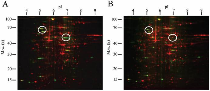
Fig. 1.

Multi-channel image of 2D electrophoresis gel of C. elegans proteins from ground control and flight experiment. Panel (A) and (B) are 2D protein mapping images of the ground control and the flight experiment, respectively. The whole lysate was extracted by protein extraction solution containing 7M urea, 2M thiourea, 4% (w/v) CHAPS, 40mM DTT. Isoelectric focusing was performed using an immobilized pH3-10 linear gradient strip (Bio-Rad). SDS-PAGE was performed using a ready made gel (PROTEAN II ReadyGel, 8-16%, Bio-Rad). Phosphoproteins (green spots) and total proteins (red spots) were detected by Pro-Q Diamond and SYPRO Ruby staining (Invitrogen), respectively. The spot patterns were analyzed using PDQuest software (Bio-Rad). Circles indicate decreasing phosphoprotein spots in flight samples.
