
Figure 8.
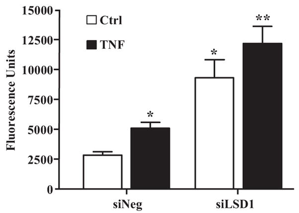
Role of LSD1 in monocyte–VSMC binding. HVSMCs were transfected with siLSD1 or siNeg, and, 48 hours later, transfected cells were transferred to serum-depletion medium. On the following day, cells were untreated or treated with TNF-α for 6 hours, and VSMC–monocyte binding assays were performed using fluorescently labeled THP-1 monocytes. VSMC monolayers were washed, and the fluorescence due to bound monocytes was quantified. Results were expressed as fluorescence units (means±SEM). *P<0.02, **P<0.014 vs siNeg (n=3).