
Figure 1. Pattern formation in the pupal eye.
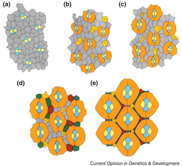
The apical surface of eyes at different stages of pupal life. Grey cells are uncommitted IPCs and coloured cells are determined. (A) At the beginning of pupation, the cone cells (blue and yellow) are embedded within a mosaic of IPCs. (B) After 20 hours pupation, PP cells (orange) are enlarging and surrounding the cone cells. (C) After 30 hours pupation, PP cells are contacting each other. (D) After 40 hours pupation, cone and PP cells are enlarging, and IPCs sort into single rows between them. IPCs positioned in certain niches differentiate into SP (green) and TP cells (red). IPCs which are not committed undergo apoptosis. (E) After 60 hours pupation, pattern formation is complete. Figure is modified after [2].