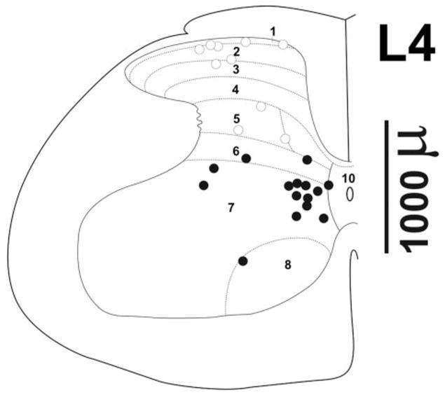
Figure 1.

Recording sites. (A) Recording sites determined from histologically verified lesions are shown overlaid on a representative drawing of the L4 lumbar spinal cord [taken from the atlas of Paxinos and Watson (15)]. Open circles: sites <1200 μm based on micrometer reading; filled circles: depths >1200 μm. The approximate recording depth can be estimated from the depth marker at right.
