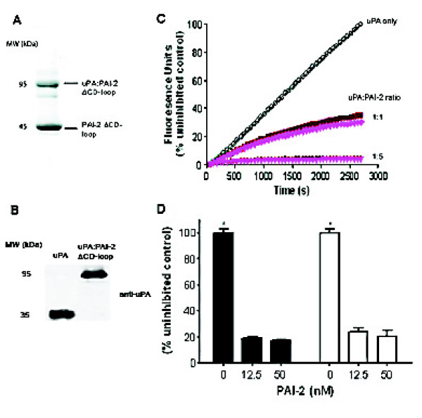
Figure 4.
PAI-2 ΔCD-loop efficiently inhibits both solution phase and cell bound uPA activity. A. PAI-2 ΔCD-loop is able to form SDS stable complexes with uPA. PAI-2 ΔCD-loop was incubated in a 2:1 molar ratio with HMW-uPA for 30 min and analysed by a 10% SDS-PAGE under reducing conditions. B. Immunoblot analysis of complex formation between uPA and PAI-2 ΔCD-loop using a monoclonal antibody to the A-chain of uPA (~30 kDa), indicating that no residual uPA remains in the sample. C. Kinetic inhibition curves for wild-type PAI-2 (dark red) versus PAI-2 ΔCD-loop (bright pink) against HMW-uPA in solution. The uPA fluorogenic substrate was briefly pre-incubated with the PAI-2 forms or buffer alone (open circles) and the assays initiated by the addition of HMW-uPA. Fluorescence units were converted to a percentage of the maximal (uninhibited) uPA activity. Values shown are the means of triplicate determinations. Errors (<10%) are not shown for clarity of presentation. D. U937 cells were incubated with uPA fluorogenic substrate in the absence or presence of wild-type PAI-2 (filled bars) or PAI-2 ΔCD-loop (open bars) for 1 h at 37°C. Fluorescence units were converted to a percentage of the maximal (uninhibited) uPA activity. Values shown are means ± SEM (n = 3). *p < 0.001 compared to inhibited samples for both PAI-2 forms.

