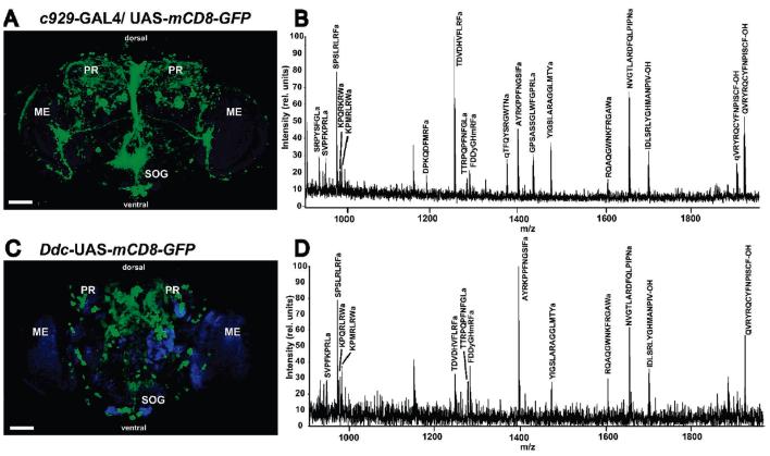
Figure 4.
Frontal confocal microscope images of D. melanogaster adult brain tissue genetically labeled with the cell-surface antigen mCD8-GFP (green) and the corresponding mass spectral profile of extract from cell populations enriched using GAL4-UAS-mediated immunoaffinity purification. Counterstaining with the anti-NC82 antibody was used to mark the neuropil regions (blue). The dimm (c929)- (A) or Ddc-GAL4 (C) driver lines targeted, respectively, primarily peptidergic or dopaminergic and serotonergic cells. Dorsal and ventral aspects of the fly brain are indicated. MALDI-TOF MS analysis of extract from neuronal subpopulations enriched using the dimm (c929)-GAL4 driver gave the broadest peptide profile (B), whereas isolation with the Ddc-GAL4 driver (D) resulted in simpler profile. Signals of identified peptide ion species have been labeled with the predicted amino acid sequence. PR, protocerebrum; ME, medulla; SOG, subesophageal ganglion. Scale bar: 50 μm.

