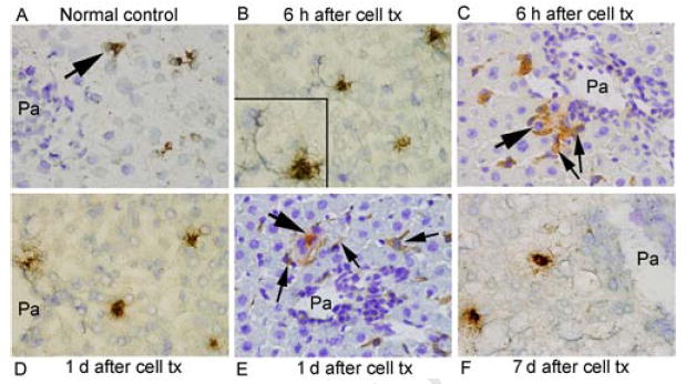
Figure 2. Immunohistological studies of PTGS1 expression.

PTGS1 in normal rat liver (A) was expressed occasionally and was more frequently expressed after cell transplantation (arrows, B-F), including HSC (inset, B). Combined staining for DPPIV and PTGS1 showed that transplanted cells (thick arrows) and cells expressing PTGS1 (thin arrows) were often in proximity to one another (C,E). Orig. mag. x400; toluidine blue counterstain.
