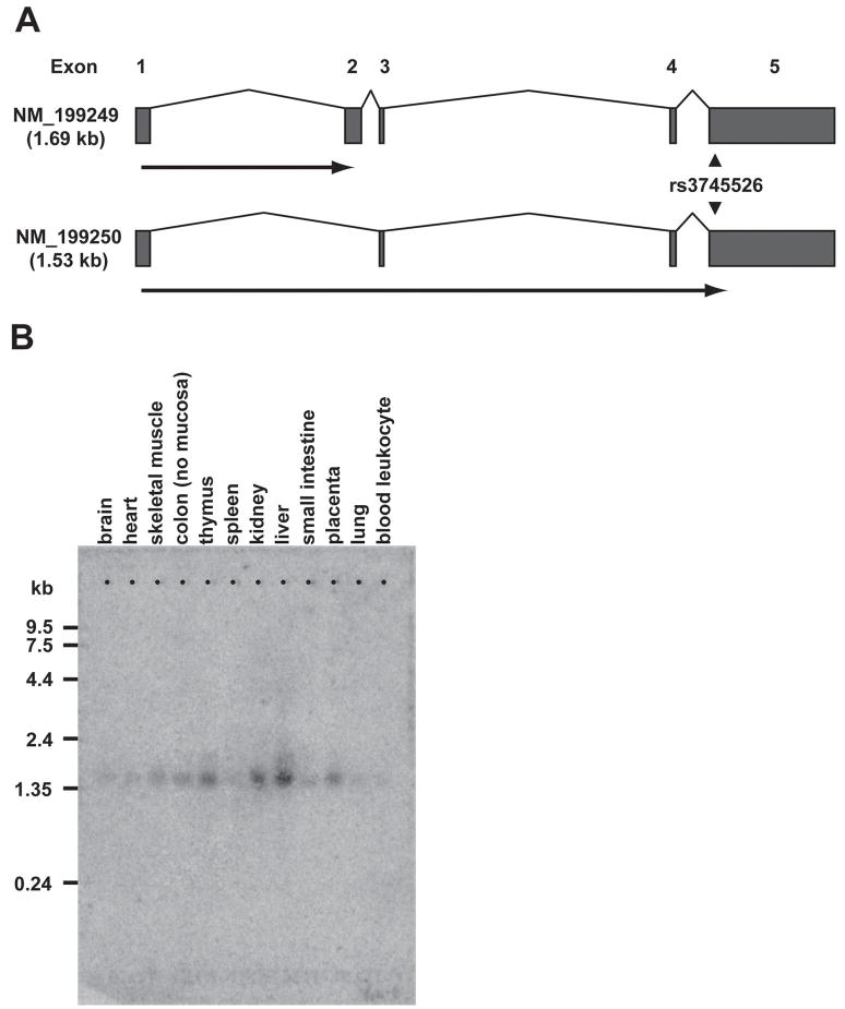
Figure 2. Two Alternative Transcripts from C19orf48.
(A) Schematic diagram of the C19orf48 genomic locus showing the intron/exon structure of the two RefSeq transcripts NM_199249 and NM_199250. The heavy horizontal arrows indicate the open reading frames (in frame +2) defined by the ATG at nucleotides 137–139 in both NM_199249 and NM_199250. The location of the nonsynonymous T↔A SNP at nucleotide 263 is noted (arrowheads). (B) An oligonucleotide spanning ORF +2/48 of the NM_199250 transcript of C19orf48 was amplified by PCR and purified (QIAGEN). This oligonucleotide was 32P-labeled using a Random Primed DNA Labeling Kit (Roche Diagnostics Corporation, Indianapolis, IN) and used to probe a Multiple Tissue Northern Blot nylon membrane (BD Biosciences/Clontech) loaded with poly (A)+ RNA isolated from 12 different human tissues. Hybridization was performed as described (41) at 68°C for one hour. Imaging was obtained by 7-day exposure on a PhosphorImager (Molecular Dynamics, Sunnyvale, CA).

