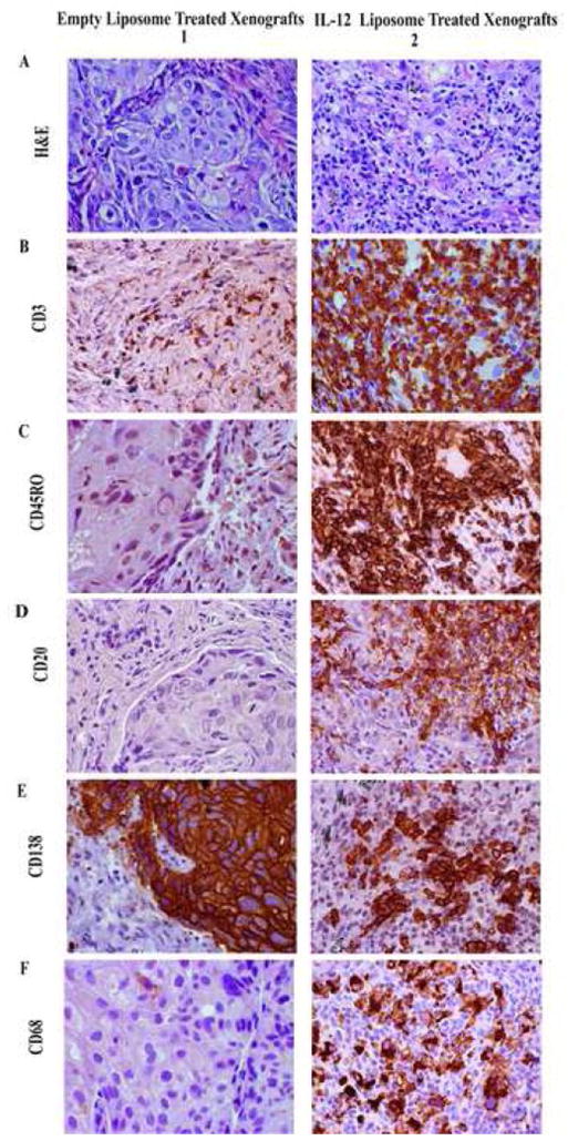
Figure 5. Immunohistochemical staining of tissues reveals that treatment of lung tumor xenografts with IL-12 liposomes promotes the expansion and survival of leukocytes within the tumor microenvironment.

Mice were implanted with a human primary lung tumor, and at 2 weeks post IL-12 or empty liposome treatment mice were sacrificed and the xenografts removed. A. H&E stain of xenografts treated with empty liposomes (1) and IL-12 liposome treated xenografts. B-C, Immunohistochemical staining revealed the presence of CD3+ CD45RO+ cells in the IL-12 liposome treated xenografts (B2-C2) but negligible staining in the control treated xenografts (B1-C1). D-F, In contrast to the diffuse accumulation of T cells throughout the IL-12 treated xenograft, fewer foci of CD20+ B cells (D2), CD138+ plasma cells (E2) and CD68+ macrophages (Fig F2) are present in the cytokine treated xenografts. Very few B cells (D1), plasma cells (E1) or macrophages (F1) are observed in any of the xenografts treated with control liposomes. All images were taken at 400X magnification.
