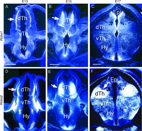
Figure 3.
Expression of Robos in embryonic rat diencephalon. Coronal sections through diencephalon of E13 (A, D), E15 (B, E), and E17 (C, F) rat brains showing Robo1 (A–C) and Robo2 (D–F) expression detected by in situ hybridization using S35 labeled riboprobes. Sections are counterstained with bisbenzimide. Photos are single exposures using dark field and ultraviolet illumination. Dorsal is up. Arrows mark Robo expression in domains of TCA projection neurons in nascent dTh nuclei. For details, see Results. dTh, dorsal thalamus; Epi, epithalamus; Hy, hypothalamus; and vTh, ventral thalamus. Scale bars: 200 mm.

