Fig. 3.
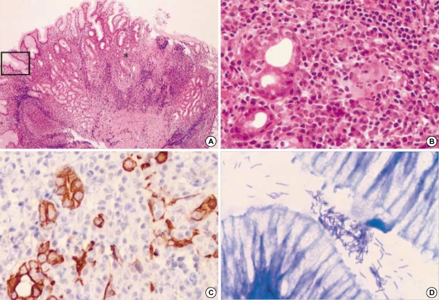
H. heilmannii-associated gastric MALT lymphoma. (A) Low magnification view reveals extensive lymphocytic infiltration (hematoxylineosin, ×100) with an asterisk indicating areas of (B) and (C), and an inset representing (D) area. (B) Lymphomatous infiltrations form destructive lymphoepithelial lesions (LELs) (hematoxylin-eosin, ×400). (C) Cytokeratin immunostain highlights the remaining epithelial remnant in LELs (×400). (D) Many spiral organisms are present in the adjacent gastric pit of this case (Giemsa stain, ×1,000).

