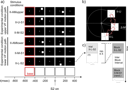
Figure 2.
Stimuli used in experiment II to test for a retinotopic modulation of the SSDWs. Each black frame corresponds to the presentation of the corresponding stimulus display for 200 ms. A red border indicates the use of this frame/time interval as a baseline interval in VEP analysis. The time axis is aligned to the onset of second stimulus in the respective AM stimulus conditions (t = 0). (a) From top to bottom: (II-AMupper) AM inducing stimulus condition with a motion path that lies predominantly in the upper visual field. (II-U-S1) Control stimulus condition that matches the first stimulus of II-AMupper in timing and location. (II-M-S2) Control stimulus condition that matches the second stimulus in II-AMupper in timing and location. (II-AMlower) AM inducing stimulus condition with a motion path that lies exclusively in the lower visual field. (II-M-S1) Control stimulus condition that matches the first stimulus of II-AMlower in timing and location. (II-L-S2) Control Stimulus condition that matches the second stimulus in II-AMlower in timing and location. (b) Stimulus geometry: all stimuli were located on a circle of an eccentricity of 4° visual angle; (II-U) first stimulus in the AM inducing condition II-AMupper, located 45° above the horizontal meridian, (II-M) second stimulus in the AM inducing condition II-AMupper or first stimulus in the AM inducing condition II-AMlower, located 15° below the horizontal meridian. (II-L) Second stimulus in the AM inducing condition II-AMlower, located 75° below the horizontal meridian. Red arrows symbolize the AM paths in the upper and lower condition. The stimulus geometry is chosen such that the representation of the lower motion path between the 2 stimuli (in condition II-AMlower) lies on the upper bank of the calcarine sulcus whereas the representation of the upper motion path between the 2 stimuli (in condition II-AMupper) lies at an opposing position on the lower bank of the calcarine sulcus. (c) Sequence of stimulus conditions in an experimental run. All stimulus condition types were presented in blocks of 6. ITIs within a block were randomized (1800–1920 ms, uniform distribution), each block of 6 was followed by a stimulus free IBI that had a randomized duration (1800–1920 ms, uniform distribution). The order of the different stimulus blocks (I-AM, I-AM(weak),…) was also randomized.

