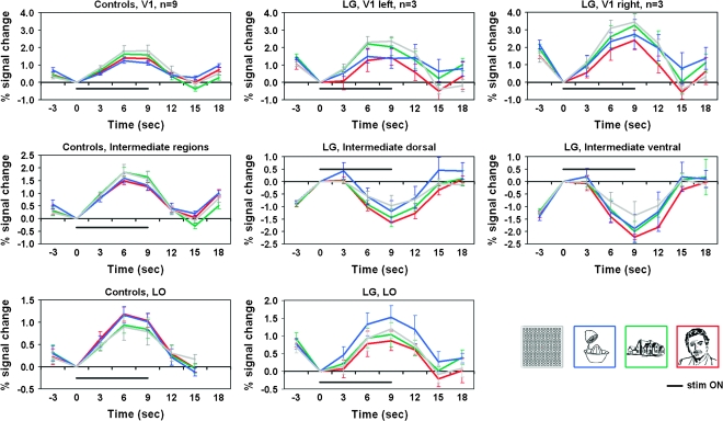
Figure 4.
Regional time courses: LG versus controls. Average time courses of the controls (n = 9) are presented in the left most column; LG's average time courses (sampled from the 3 category-localizer runs he underwent) are presented in the middlle and right columns. The V1 average time courses are displayed in the top row (LG's right and left hemispheres are presented seperately since the right hemisphere was significantly different from both the left hemisphere and from the controls (see Results for details). V1 sampled regions were determined for LG as well as for controls according to activated patches within V1 retinotopic boundaries. Time courses sampled from intermediate-level regions are displayed in the middle row (LG's ventral and dorsal aspects are presented seperately since a significant difference was found between them; no inter-hemisphereic difference was found). Intermediate level regions were determined for controls based on retinotpic borders, so that the sampling was based on activated patches within V2–V3 regions. For LG, intermediate regions were sampled from the deactivated patches in the corresponding anatomical location of V2–V3 in controls. LO related time courses (sampled from the visually activated regions in the anatomical location of LO for both LG and controls) are displayed in the bottom row. The average response to the faces condition is indicated by the red curve, to houses in green, to objects in blue, and to the patterns condition in gray. Stimulus “ON” is indicated by a black line. Error bars denote SEM. Note the prominent deactivation to each of the categories in LG's intermediate areas (middle row). For more details see Methods and Results.

