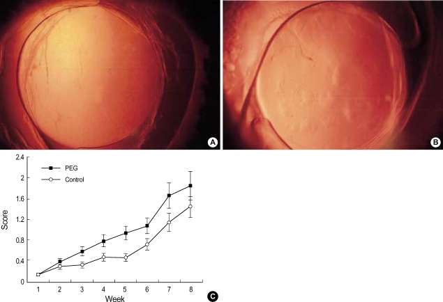
Fig. 3.
The formation of posterior capsular opacification (PCO) after implantation of intraocular lens in rabbits. (A) Polyethylene glycol (PEG), (B) Control. At the 3rd week, the fine fibrotic materials were found on the posterior capsule with minimal proliferation of lens epithelial cells (LECs) in comparison with the control, where the conglomerated LEC's were dominant. (C) The severity of PCO was significantly lower from the third week in the PEG group until the sixth week of examination. *, p<0.05 by Mann Whitney-U test.

