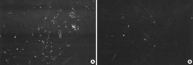
Fig. 5.
The scanning electron microscope of adherent cells (×200 fold). (A) In the control group, spindle shape, patch-like cells, together with a few small round cells, were firmly attached to lens surface. (B) In the polyethylence glycol (PEG) group similar cells were found but seemed less adherent.

