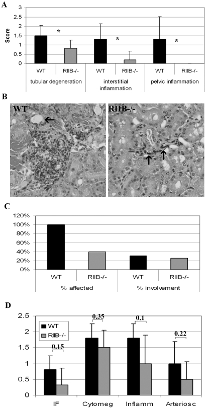
Figure 8. Old RIIβ−/− male mice show attenuated renal dysfunction, tumor incidence, and a trend towards attenuated cardiac pathology.
A. Old RIIβ−/− male mice showed attenuated tubular degeneration and interstitial and pelvic inflammation compared to wild-type littermates although renal lesions in both genotypes was relatively mild. N = 6. Numbers represent means. Error bars represent standard deviations. *P<0.05 (determined by Student's t-test). B. Hematoxylin and eosin-stained section of kidneys from WT and RIIβ−/− male mice. The WT mouse had mild perivascular and interstitial lymphohistiocytic accumulations (severity score 2) and mild tubular changes including attenuated epithelium and intralumenal proteinaceous material (arrow). This mouse also had marked neutrophilic pyleitis that likely contributed to moribundity. In contrast, the RIIβ−/− mouse had minimal interstitial inflammation (severity score 1) surrounding a small caliper tangentially sectioned vessel (arrows). Original magnification for both images, 20X. C. Spleens from 100% of WT mice but only 40% of RIIβ−/− mice examined at end of life, presented with lesions. N = 6. D. Hearts from old (18 month) RIIβ−/− male mice tended towards better scores than WT for 4 of 6 types of lesions graded, although differences were not significant. IR = interstitial fibrosis, cytomeg = cytomegaly, inflamm = inflammation, arteriosc = arteriosclerosis. N = 5 (WT) and 6 (RIIβ−/−). Numbers represent means. Error bars represent standard deviations. Numbers above columns represent probability values, determined by Student's t-test.

