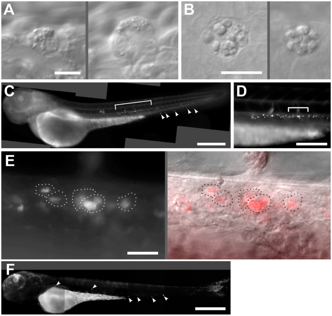
Figure 2. Leptospirosis of the zebrafish embryo at 24 hours post infection.
A. Two affected cells in the caudal vein containing cytoplamsic vesicles, now larger. This embryo was infected intavenously. Scale bar, 10 µm. B. Affected cells in the brain, apparently containing clusters of undigested apoptotic bodies. This embryo was infected via hindbrain ventricle. Scale bar, 10 µm. C. Fluorescent image of whole embryo infected intravenously with SYTO 83-stained leptospira. While some fluorescent leptospires appear around the ventral tail (arrowheads), the majority have localized near the dorsal aorta (bracket). Scale bar, 300 µm. D. Higher magnification of the area bracketed in E, showing numerous distinct clusters of stained leptospires lateral to the dorsal aorta, just ventral to the notochord. Scale bar, 100 µm. E. Higher magnification of the area bracketed in D, with SYTO 83 fluorescence to the left and DIC overlay to the right. See Video S5. Dotted lines indicate the outlines of infected cells. Scale bar 20 µm. F. Fluorescence image of embryo 24 hours after infection with green fluorescent P. aeruginosa. Infected cells (arrowheads) appear in various places throughout the circulation. Scale bar 300 µm.

