
Figure 6.
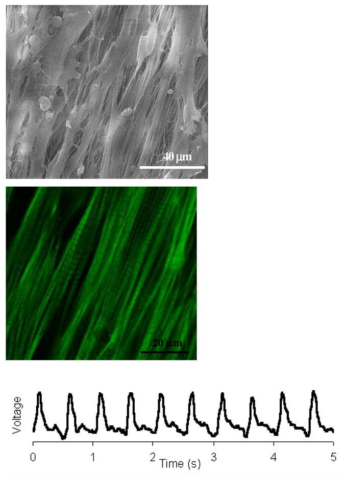
(a) SEM images of cardiac myocytes cultured on uniaxially stretched aligned PLLA electrospun scaffolds, (b) corresponding confocal micrograph of (a); (c) electrical response of cardiac myocytes on electrospun scaffolds (action potentials were measured using a voltage-sensitive dye di-8-ANEPPS and a micro scale optical recording system). (From Ref. [133] with permission)