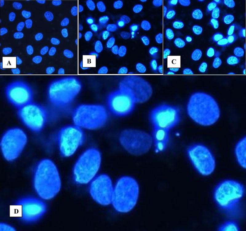
Figure 1.

Cells in culture examined by fluorescence microscopy following staining with 4′–6-diamidino-2-phenylinedole. A. Normal NRK52E cells showing normal nuclei. Reduced from X20. B, C. Cells exposed to HA for 3 and 6 hours show condensed chromatin. Reduced from X20. D. High mag of Fig 1B showing both normal appearing nuclei as well as nuclei with condensed chromatin, and formation of apoptotic bodies. Reduced from X40.
