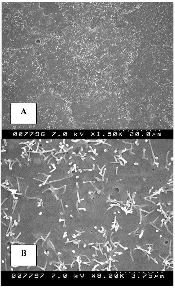
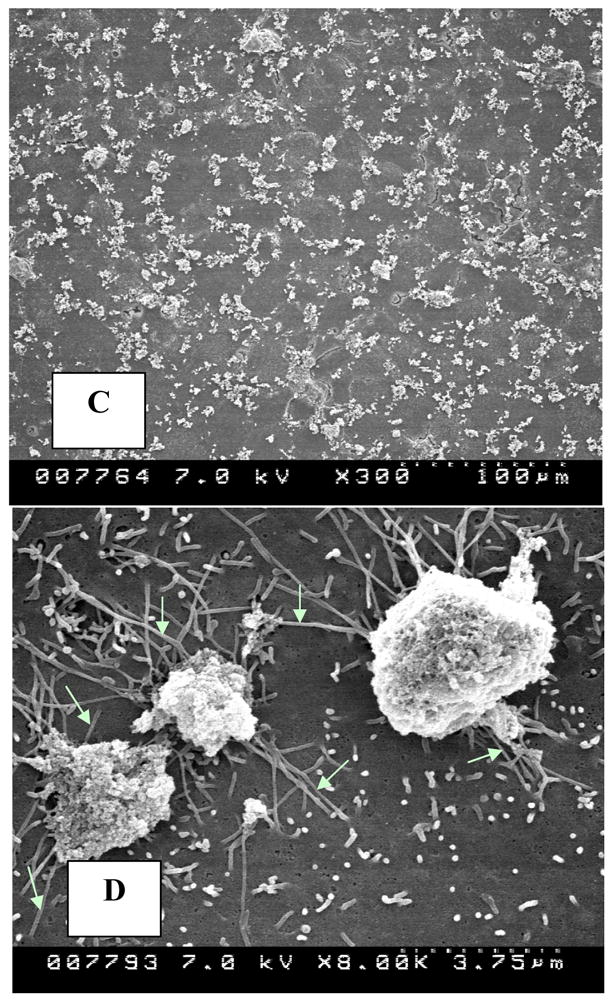
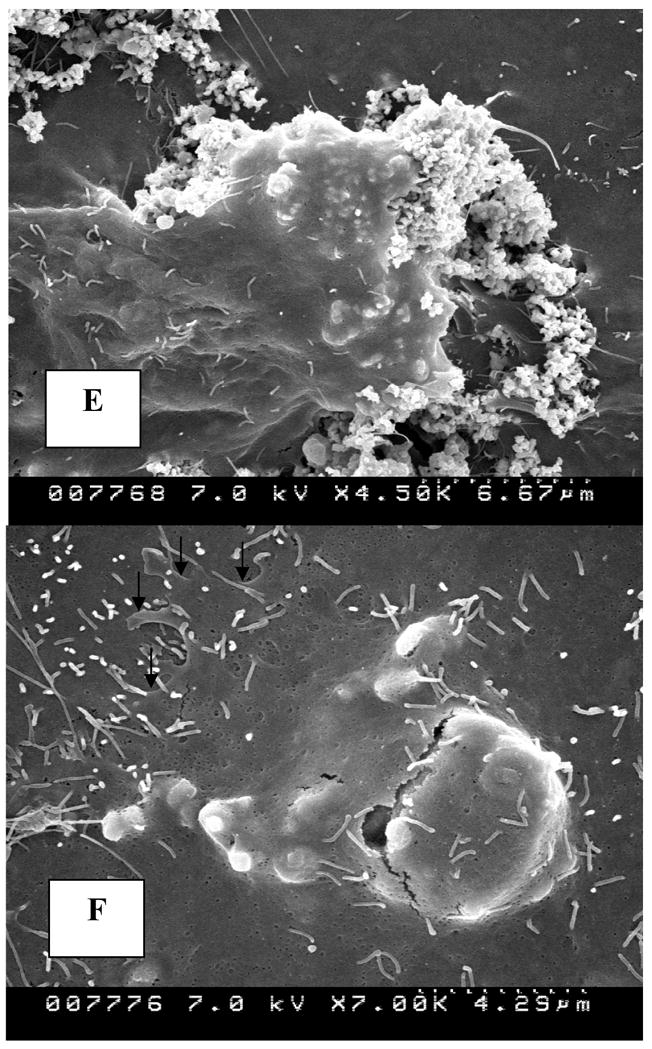
Figure 2.


Cells in culture examined by scanning electron microscopy. A, B. Normal epithelial cells showing squamous epithelial surface morphology with sparse stubby microvilli. C. Cells exposed to 133μg/cm2 HA show crystal clumps distributed on cell surface. D. Slender extensions of cells appear to probe crystal clumps (white arrows). E. A cell is endocytosing a crystal clump with cell margin growing over the crystals. F. Crystal is endocytosed. Edge of the endocytosing cell (arrows) can still be distinguished from the underlying cell. (Magnifications-Figure 2A X1500, 2B X8000, 2C X300, 2D X8000, 2E X4500K, 2F X7000)
