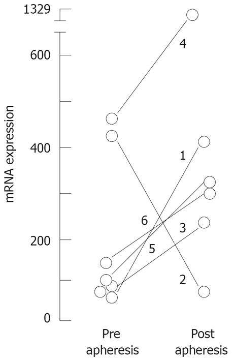
Figure 2.

Variation of mRNA expression for FoxP3 in CD4+ T cells in matched samples from 6 patients treated by GMA-apheresis: Quantitative PCR was performed as described in methods; Results are expressed as FoxP3 expression relative to GUS expression per cent.
