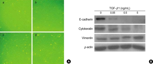
Fig. 1.
The concentration of TGF-β1 to induce A549 cells to undergo EMT. A549 cells were incubated with TGF-β1 at various concentrations for 48 hr. (A) Morphologic changes are observed with 0.05 ng/mL of TGF-β1, and TGF-β1 treated cells with concentration as high as 5 ng/mL show more fibroblast-like morphologic changes. a, control; b, 0.05 ng/mL; c, 0.5 ng/mL; d, 5 ng/mL TGF-β1. (B) Expression of epithelial marker E-cadherin and cytokeratin are down-regulated by TGF-β1 stimulation in a concentration-dependent manner.

