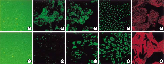
Fig. 2.
Epithelial-to-mesenchymal transition of A549 cells in vitro. A549 cells treated without (upper column) or with 5 ng/mL TGF-β1 (lower column) for 48 hr in serum-free medium. A549 cells changed to more elongated, fibroblast-like cells (A, F). Mesenchymal transition cells revealed loss of E-cadherin (B, G), cytokeratin (C, H), cytokeratin replacement by vimentin (D, I), and stress fiber reorganization by F-actin (E, F).

