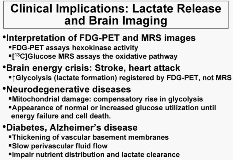
Fig. 6. Imaging neurodegenerative diseases.
Slow, progressive evolution of mitochondrial damage may lead to compensatory increases in glycolysis. Increased glycolytic utilization of glucose would be registered by [18F]fluorodeoxyglucose (FDG)-positron emission tomographic (PET) studies, but not by use of labeled glucose if lactate efflux from brain is increased. Glucose utilization rates that are within or above the normal range during early, potentially-treatable stages of disease may not be appropriately interpreted unless lactate generation and release is also assessed when imaging of diseased brain is carried out. Pathophysiological changes involving the vasculature could affect nutrient and by-product trafficking, as well as transport of tracers used for brain imaging and clearance of their metabolites.

