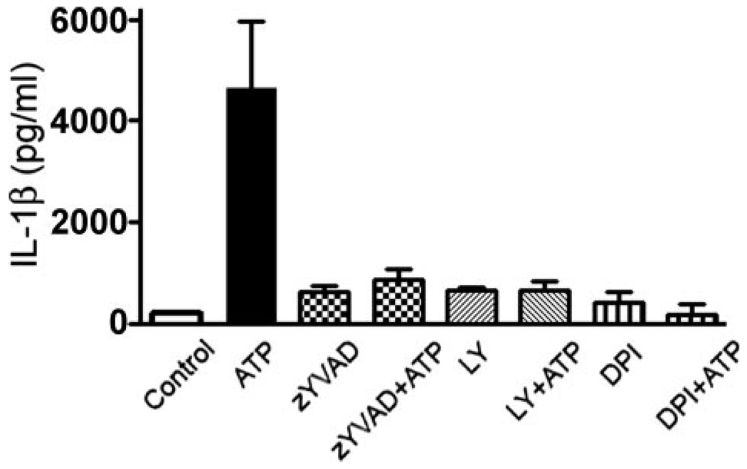
FIGURE 8. ATP stimulation of macrophages leads to IL-1β secretion through a pathway requiring ROS production and PI3K activation.
Macrophages were primed with 1 µg/ml LPS for 2 h at 37 °C, before treating the macrophages with the caspase-1 inhibitor, Z-YVAD-fmk (50 µm) for 30 min, DPI (2 µm) for 10 min, or LY294002 (50 µm) for 10 min. The cells were then stimulated with 3 mm ATP for 6 h. Secretion of IL-1β was measured by ELISA. p < 0.001 for cells treated with ATP and Z-YVAD-fmk, DPI, or LY294002, compared with cells treated with ATP alone. The experiment was performed twice in duplicate, and the results represent the average and S.D.

