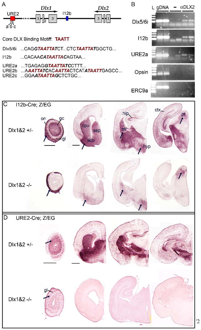
Figure 12. DLX2 binds directly to I12b and URE2 in vivo and DLX1&2 function is necessary for I12b and URE2 activity.

(A) Schematic of the Dlx1 and Dlx2 genomic locus showing the location of I12b and URE2 regulatory elements; the three putative DLX binding sites within URE2 are labeled (a-c). The core binding motif for DLX transcription factors is predicted to be TAATT. Dlx5/6i is a regulatory element in the intergenic region between Dlx5 and Dlx6 known to bind DLX1 and DLX2 in vivo. The DLX binding motif is present in Dlx5/6i, I12b, and URE2 enhancer elements (red italics).
(B) Chromatin immunoprecipitation (ChIP) assays from E15.5 ganglion eminences using DLX2-specific antisera (aDLX2) shows that DLX2 binds directly to Dlx5/6i (top panel) and also to I12b (second row) and URE2 (third row) in vivo. Control assays without antibody or with DLX2 antisera followed by PCR to irrelevant genomic loci (Opsin promoter region or ERC9a, an enhancer region upstream of the progesterone receptor), were negative. L, 100 bp DNA ladder; gDNA, genomic DNA; -, no antibody.
(C) I12b activity was greatly diminished in Dlx1&2 mutants (Dlx1&2-/-) with some notable exceptions (arrows). I12b-Cre;Z/EG animals were bred with Dlx1&2 mutant mice to generate Dlx1&2 null mutants that were transgenic for I12b-Cre and Z/EG. EGFP expression was visualized in coronal brain sections from control (top row) or Dlx1&2-/- mutants (bottom row) at P0.
(D) URE2 activity is nearly abolished in Dlx1&2 mutants. URE2-Cre;Z/EG animals were bred with Dlx1&2 mutant mice to generate Dlx1&2 null mutants that were transgenic for URE2-Cre and the EGFP reporter. EGFP expression was visualized in coronal forebrain sections from control (top row) or DLX1&2-/- mutants (bottom row) at P0. Expression was lost throughout the brain except in a few cells within the glomerular layer of the olfactory bulb (arrows).
acb, accumbens; cpu, caudate-putamen; ctx, cortex; gc, granule cell layer; gl, glomerular layer; hip, hippocampus; hyp, hypothalamus; on, olfactory nerve layer; pt, pretectal nucleus; sep, septum.
Scale bars = 500 μm.
