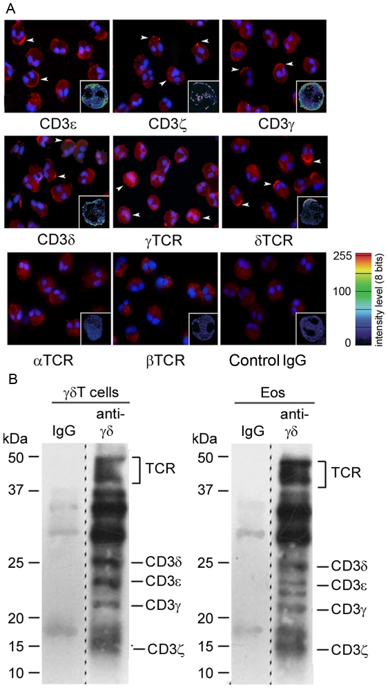
Figure 4. Immunolocalization and Subunit composition of surface-expressed γδTCR/CD3 complex on human eosinophils.
(A) Immunofluorescence and confocal microscopy (insets) analysis of γδTCR/CD3 chains expression on cytospin eosinophil preparations after 18 h culture. Staining with anti-αTCR, anti-βTCR antibodies and control goat IgG is represented. Arrowheads indicate some positive cells. (B) Surface of eosinophils and γδT cells was biotin-labelled. Cells were lysed and complexes were immunoprecipitated, using anti-γδTCR or isotype control antibodies. Immunoprecipitated proteins were resolved on a reducing 14% SDS-PAGE and transferred to PVDF membrane. Biotinylated proteins were revealed using ABC-HRP and chemiluminescence. Positions of the TCR and CD3 subunits are marked. Material corresponding to the total and to 1/10th of the material was loaded for eosinophils and γδT cells respectively. Dashed lines indicate that non adjacent lanes of the same gel have been joined on the Figure.

