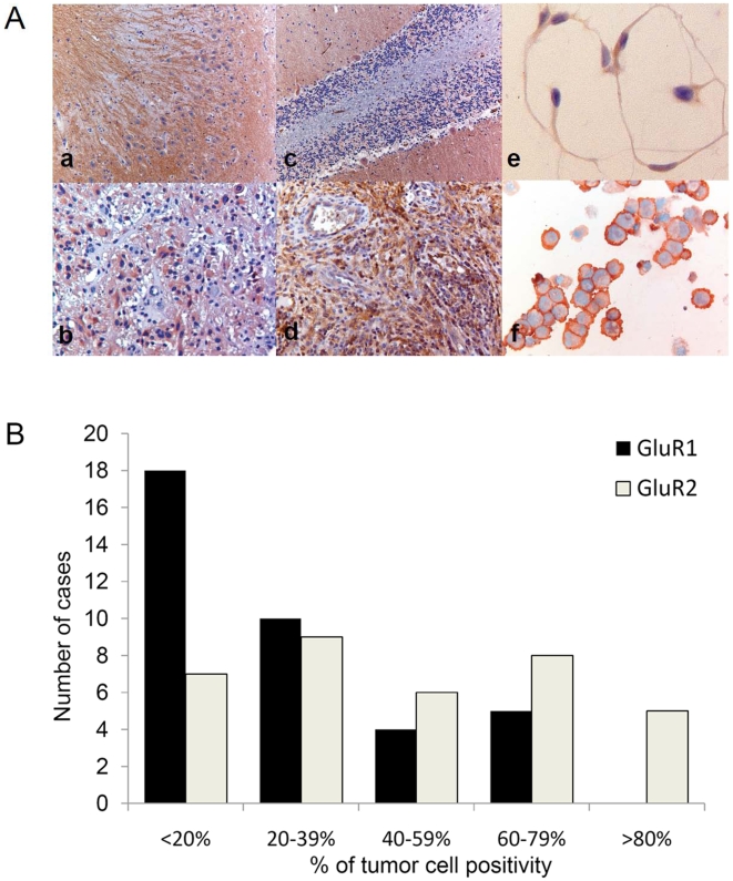
Figure 2. AMPAR immunohistochemistry on paraffin embedded sections.
(A) Representative images of (a) GluR1 in hippocampus, (b) GluR1 in GBM, (c) GluR2 in cerebellum, (d) GluR2 in GBM, (e) GluR2 staining in chamberslide of VU-122 cell line, (f) GluR2 staining in cytospin of VU-122. (B) Histogram of protein expression of GluR1 and GluR2 in 37 cases of adult GBM.

