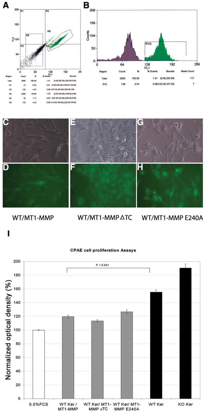
Figure 3.

Effect of keratocyte MT1-MMP overexpression on CPAE proliferation. WT keratocytes transfected with MT1-MMP were sorted by flow cytometry based on GFP expression (A, B). Flow cytometry was also used to monitor the stability of GFP expression. Fluorescence microscopy was used to confirm expression of MT1-MMP (C, D), MT1-MMP-ΔTC (E, F), and MT1-MMP-E240A (G, H). CPAE cells were incubated for 48 h with conditioned media from WT keratocytes stably transfected with WT or mutant MT1-MMP. The colored formazan product assay was then performed to measure CPAE cell proliferation (I). Experiments were performed using 0.5% FCS as a negative control and 10% FCS as a positive control. Optical densities (OD) were normalized relative to the 0.5% FCS OD, which was set at 100%. Proliferation values obtained in the presence of WT and KO conditioned media from Fig. 1 are included for comparison (solid black bars).
